JoAnn Pushkin Breast Cancer
Name of patient
Disease type

Personal story
JoAnn Pushkin detected her own breast cancer when she was 45 years old. JoAnn lives in the U.S. and though she was a diligent patient and never missed an annual mammogram, did self-exams, ate healthy and had no known risk factors for breast cancer, one day she felt a lump.
Having just had a recent “normal” mammogram, she wasn’t particularly concerned, but contacted her doctor as a precaution. She was immediately sent for a diagnostic mammogram to be followed by an ultrasound. To her confusion and shock, the diagnostic mammogram came back “clear” and did not show the cancer that was large enough to feel. It was the ultrasound that followed that revealed the sad news that she had breast cancer. By the size, the cancer was estimated to have been growing for up to 5 years in her breast – undetected every one of those years by mammography. On the same day that JoAnn learned she had breast cancer, she also learned she had extremely dense breast tissue and that her dense tissue was the reason the cancer was obscured on her mammograms. She had never been told she had dense breasts, never told that breast density increased her risk for breast cancer, never told that breast density can drastically compromise the effectiveness of a mammogram, and, unfortunately, never told that additional screening tools were available that might have detected her cancer at an earlier stage.
JoAnn Pushkin’s story and advocacy was the inspiration for New York State’s Dense Breast Inform law; she was also instrumental in the introduction of U.S. Federal regulatory and legislative efforts for patient “dense breast” notification. To address the need for education on the topic, she co-founded and is Executive Director of the world’s leading website on the topic, DenseBreast-info.org.
Radiological Findings
Why a Conversation About Your Breast Density Matters
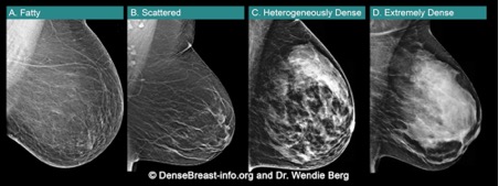
Breasts are made of fat and glands (that make milk) held together by fibrous tissue. The more glands and fibrous tissue a woman has, the “denser” her breast tissue.
• Breast density is seen on a mammogram
and described as one of four categories. Fig.1
(A) Fatty;
(B) Scattered fibroglandular density;
(C) Heterogeneously dense; or
(D) Extremely dense
(C) Heterogeneously dense or (D) Extremely dense are considered “dense breasts.”
• Dense breasts are normal. Almost half of women over age 40 have dense breasts.
• Although normal, dense breasts make it harder to see cancer on a mammogram. Cancers can be hidden and missed until they are larger and more likely to have spread.
• Dense breasts increase the risk of getting breast cancer. The denser the breasts, the greater the risk.
• After a mammogram, other screening tests, such as MRI or Ultrasound, find more early-stage cancers in dense breasts.*
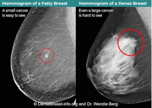
Fig.2 shows cancer on a mammogram of a fatty breast vs. a dense breast.
Talk to your doctor or health professional about what additional screening tests may be recommended for you based on your risk factors, including breast density.
*EUSOBI guidelines now recommend MRI every 2 to 4 years after a mammogram in all women with extremely dense breasts. If MRI is not an option, Ultrasound can be used. DenseBreast-info.org | © 2015-2022
For more information, visit: DenseBreast-info.org/Europe
References
- Resources:
- DenseBreast-info.org/Europe
- Additional Reading:
- Breast density implications and supplemental screening, Athina Vourtsis, Wendie A Berg, European Radiology (2019) 29:1762-1777
- Using education to overcome unequal access to supplemental screening for women with dense breasts, Dr. A.Vourtsis, Dr. W.A.Berg, Diagnostic Imaging Europe (2020)