Clare Cowhig Breast Cancer
Name of patient
Disease type

Personal story
Clare lives in the UK. Due to her significant family history, Clare began mammography screening at 41. In April 2018, aged 50, Clare was shocked to be diagnosed with bilateral breast cancer despite having “normal” annual mammograms, which had always been reported as clear.
Clare had presumed all was well. However, 9 months after her last annual mammogram she felt an unusual, thickened area on her breast. Thankfully, she decided to go for private whole breast ultrasounds. The sonographer informed Clare that she had the densest breast tissue she’d ever seen. In her opinion Clare should have been having MRIs.
DIAGNOSIS: An MRI scan and core biopsies confirmed a Stage 3 Invasive Ductal Carcinoma over 5cms on the right and a Stage 2, Invasive Ductal Carcinoma of 2cms on the left with additional areas of DCIS in both.
Despite 2 surgeries, more cancer was found so after 5 months of chemotherapy Clare had a double mastectomy. According to Clare “I kept thinking that this may have been avoided if I had been informed that I was at very high risk of false negative results. I could have discussed this with my team and reached some shared decisions about additional screening.”
If Clare had relied solely on yearly mammograms, it is scary to think how advanced her cancer could have become.
Clare believes earlier detection would have saved her and her family a whole lot of pain and anxiety.
Clare now works to help raise awareness. All women need to know that dense breast tissue may require additional screening and can also increase your risk of developing breast cancer. This is such an important issue. Knowledge is Power!
Until there’s a cure #FindItEarly
Watch my Dense Breast story: Dense Breast Diaries – Clare’s Story (UK)
Radiological Findings
Why a Conversation About Your Breast Density Matters
Breasts are made of fat and glands (that make milk) held together by fibrous tissue. The more glands and fibrous tissue a woman has, the “denser” her breast tissue.
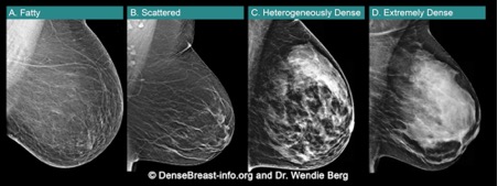
• Breast density is seen on a mammogram
and described as one of four categories. Fig.1
(A) Fatty;
(B) Scattered fibroglandular density;
(C) Heterogeneously dense; or
(D) Extremely dense
(C) Heterogeneously dense or (D) Extremely dense are considered “dense breasts.”
• Dense breasts are normal. Almost half of women over age 40 have dense breasts.
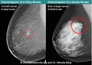
• Although normal, dense breasts make it harder to see cancer on a mammogram. Cancers can be hidden and missed until they are larger and more likely to have spread.
• Dense breasts increase the risk of getting breast cancer. The denser the breasts, the greater the risk.
• After a mammogram, other screening tests, such as MRI or Ultrasound, find more early-stage cancers in dense breasts.*
Fig.2 shows cancer on a mammogram of a fatty breast vs. a dense breast.
Talk to your doctor or health professional about what additional screening tests may be recommended for you based on your risk factors, including breast density.
*EUSOBI guidelines now recommend MRI every 2 to 4 years after a mammogram in all women with extremely dense breasts. If MRI is not an option, Ultrasound can be used. DenseBreast-info.org | © 2015-2022
For more information, visit: DenseBreast-info.org/Europe
References
- Resources:
- DenseBreast-info.org/Europe
- Additional Reading:
- Breast density implications and supplemental screening, Athina Vourtsis, Wendie A Berg, European Radiology (2019) 29:1762-1777
- Using education to overcome unequal access to supplemental screening for women with dense breasts, Dr. A.Vourtsis, Dr. W.A.Berg, Diagnostic Imaging Europe (2020)