Cheryl Cruwys Breast Cancer
Name of patient
Disease type

Personal story
Cheryl lives in France and in March 2016, aged 50, she was invited for her first ever mammogram. The radiologist announced it was ‘normal’; no abnormalities, but a decision was made to carry out an ultrasound as there was an area of dense tissue. Cheryl later discovered the reason for the additional screening was due to her dense breast tissue, as in France it is standard to offer ultrasound screening after a mammogram. This is because cancers are difficult to see, and sometimes hidden in dense breast tissue. Indeed, the ultrasound showed an area of concern that was not seen on mammography and the following week a biopsy confirmed a small 8-mm, grade 1 invasive cancerous tumour.
A lumpectomy was performed to remove the tumour, and Cheryl received several weeks of radiotherapy treatment. Her tumour was detected early. She did not need a mastectomy, reconstructive surgery or chemotherapy.
The French breast screening programme protocol, offering additional ultrasound screening in women with dense breast tissue contributed to her positive health outcome. Cheryl is originally from the UK and noted, “I had no symptoms and, without a doubt, if I was living in the UK, and had received a routine mammogram only, my cancer would have been left undetected based on existing UK national breast screening guidelines.”
Following her experience and positive outcome in France, Cheryl was compelled to raise awareness. She formed Breast Density Matters UK, a patient advocacy group, with the aim to educate UK women about dense breast implications. Equipped with breast density information, women can have informed conversations and make shared decisions about their breast health discussing screening options with medical experts. Cheryl is also Education Coordinator for DenseBreast-info.org, the world’s leading medically sourced website on the topic of dense breasts.
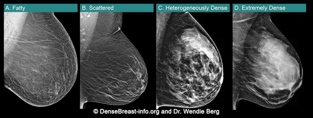
Figure 1
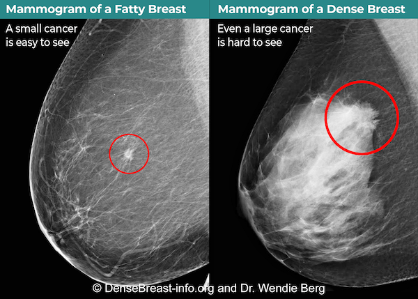
Figure 2
Radiological Findings
Why a Conversation About Your Breast Density Matters
Breasts are made of fat and glands (that make milk) held together by fibrous tissue. The more glands and fibrous tissue a woman has, the “denser” her breast tissue.
• Breast density is seen on a mammogram and described as one of four categories. See Fig.1
(A) Fatty;
(B) Scattered fibroglandular density;
(C) Heterogeneously dense; or
(D) Extremely dense
(C) Heterogeneously dense or (D) Extremely dense are considered “dense breasts.”
• Dense breasts are normal. Almost half of women over age 40 have dense breasts.
• Although normal, dense breasts make it harder to see cancer on a mammogram.
Cancers can be hidden and missed until they are larger and more likely to have spread.
• Dense breasts increase the risk of getting breast cancer. The denser the breasts, the greater the risk.
• After a mammogram, other screening tests, such as MRI or Ultrasound, find more early-stage cancers in dense breasts.*
Fig.2 shows cancer on a mammogram of a fatty breast vs. a dense breast.
Talk to your doctor or health professional about what additional screening tests may be recommended for you based on your risk factors, including breast density.
*EUSOBI guidelines now recommend MRI every 2 to 4 years after a mammogram in all women with extremely dense breasts. If MRI is not an option, ultrasound can be used. DenseBreast-info.org | © 2015-2022
For more information, visit: DenseBreast-info.org/Europe
References
- Resources:
- DenseBreast-info.org/Europe
- Additional Reading:
- Breast density implications and supplemental screening, Athina Vourtsis, Wendie A Berg, European Radiology (2019) 29:1762-1777
- Using education to overcome unequal access to supplemental screening for women with dense breasts, Dr. A.Vourtsis, Dr. W.A.Berg, Diagnostic Imaging Europe (2020)