Mrs B. Endometriosis
Name of patient
Disease type

Personal story
Mrs B experienced severe dysmenorrhoea from age 17 onwards. This was debilitating for a few days each month.
At age 25, a very large cyst was identified on ultrasound. Surgery revealed a 12 cm Endometrioma (chocolate cyst), the left ovary and fallopian tube were removed by laparotomy and posterior cervix disease cauterized. A successful pregnancy followed a miscarriage. Her daughter was born but severe dysmenorrhoea recurred after breastfeeding ceased.
In subsequent years, multiple cysts appeared in the remaining right ovary. The debilitating pain changed in nature and became constant. From 1999, she had a frozen pelvis, multiple laparoscopic surgeries to remove the appendix, deep rectovaginal nodule 1.5 cm and ureteral endometriosis all demonstrated by pathology. Following this, she is unable to sit/stand/function and had to lie down 24/7 for many years due to the severity of the pain. She lost all muscle strength and had constant neuropathic pain due to nerve damage/involvement.
Diffuse Adenomyosis and fibroids were identified on MRI/ultrasound.
Now aged 67, an additional rectovaginal endometriotic nodule has been identified on ultrasound. Her persistent pain continues and she has not been able to work for the past 22 years. She is able to function but with much reduced activity levels. This has been a difficult burden for her both physically and emotionally for 45+ years. It has also had a major impact on her family over a very long period of time affecting all aspects of their lives.
Radiological Findings
Endometriosis is a common benign female pathology (prevalence of 5-20%) defined by the migration of uterine tissues (endometrium) out of uterine cavity, mostly into the pelvic cavity (1); these tissues respond to hormonal stimulations of the woman reproductive age and cause various symptoms (chronic pelvic pain, infertility, dysmenorrhea, dyschesia, dysuria e.g.). Extra-pelvic localisations of the disease are rarer (digestive tract, urinary tract, umbilicus and abdominal wall, liver, pancreas, breast, nerves e.g.) (2).
The three following figures illustrate pelvic disorders due to a case of endometriosis. MRI provides a comprehensive assessment of pelvic endometriosis.
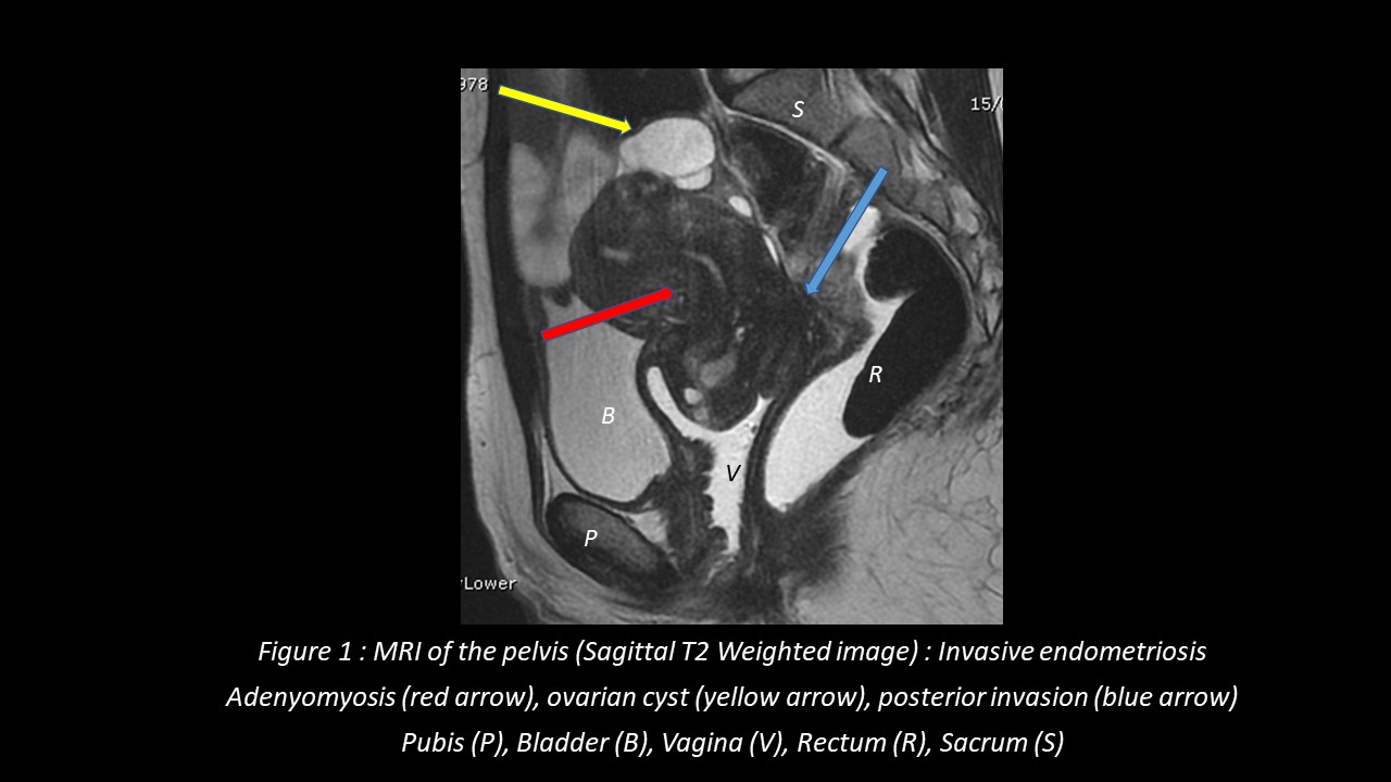
Figure 1:
Here (Sagittal T2 weighted image), the uterus shows signs of adenomyosis (abnormal endometrial implants in the uterine muscle/myometrium). Endometriosis has caused ovarian cysts and fibrotic implants at the back of the pelvic cavity, between the uterus and rectum.
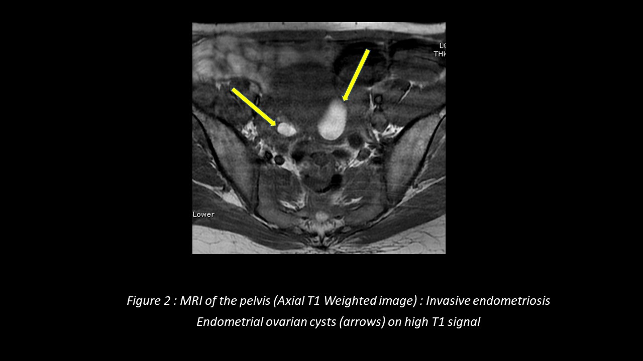
Figure 2:
On this axial MRI (in T1 weighted image) 2 endometriotic cysts developed on the ovaries appear in hypersignal (white) because they consist of blood residues.
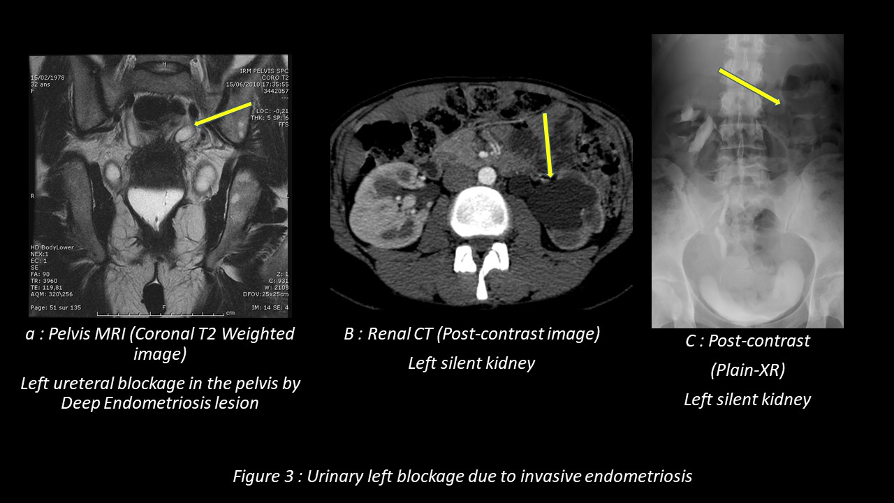
Figure 3:
The ureters are 2 thick tubes which act to transport urine from the 2 kidneys to the bladder. Endometriosis can block these tubes and cause stagnation of urine made by the kidneys. Here on this coronal MRI (in T2 weighted image) the pelvic endometriosis implant behind the bladder blocks the junction between the left ureter and the bladder (figure 3a). The contrast medium used on the CT scan (figure 3b) can no longer be excreted in the urine by the left kidney, which explains why the left kidney is called “silent”(figure 3c).
References
- Endometriosis: clinical features, MR imaging findings and pathologic correlation
Pietro Valerio Foti1, Renato Farina2 , Stefano Palmucci2, Ilenia Anna Agata Vizzini2, Norma Libertini2, Maria Coronella2, Saveria Spadola3, Rosario Caltabiano3, Marco Iraci4, Antonio Basile2, Pietro Milone2, Antonio Cianci4, Giovanni Carlo Ettorre2
2018 Apr;9(2):149-172.
doi: 10.1007/s13244-017-0591-0. Epub 2018 Feb 15. Insights Imaging - Magnetic Resonance Imaging in endometriosis-associated pain
Veronica Celli1, Sandra Ciulla1, Miriam Dolciami1, Serena Satta1, Giada Ercolani1, Maria G Porpora2, Carlo Catalano1, Lucia Manganaro3 2021 Oct;73(5):553-571. doi: 10.23736/S2724-606X.21.04782-1. Epub 2021 Apr 27 Minerva Obstet Gynecol - .MRI in female pelvis: an ESUR/ESR survey
Stephanie Nougaret1 2, Yulia Lakhman3, Sophie Gourgou4, Rahel Kubik-Huch5, Lorenzo Derchi6, Evis Sala7, Rosemarie Forstner8, European Society of Radiology (ESR) and the European Society of Urogenital Radiology (ESUR)
2022 Mar 28;13(1):60.
doi: 10.1186/s13244-021-01152-w Insights Imaging - European society of urogenital radiology (ESUR) guidelines: MR imaging of pelvic endometriosis
M Bazot1, N Bharwani2, C Huchon3, K Kinkel4, T M Cunha5, A Guerra6, L Manganaro7, L Buñesch8, A Kido9, K Togashi9, I Thomassin-Naggara10, A G Rockall11 2017 Jul;27(7):2765-2775. doi: 10.1007/s00330-016-4673-z. Epub 2016 Dec 5. Eur Radiol